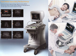
ABDOMEN UTRASOUND
An abdominal ultrasound uses reflected sound waves to produce a picture of the organs and other structures in the upper abdomen. Occasionally a specialized ultrasound is ordered for a detailed evaluation of a specific organ, such as a kidney ultrasound. An abdominal ultrasound can evaluate the:
»
Abdominal aorta, which is the large blood vessel (artery) that passes down the back of the chest and abdomen. The aorta supplies blood to the lower part of the body and the legs.
»
Liver, which is a large dome-shaped organ that lies under the rib cage on the right side of the abdomen. The liver produces bile (a substance that helps digest fat), stores sugars, and breaks down many of the body's waste products.
»
Gallbladder, which is a saclike organ beneath the liver. The gallbladder stores bile. When food is eaten, the gallblad der contracts, sending bile into the intestines to help in digesting food and absorbing fat-soluble vitamins.
»
Spleen, which is the soft, round organ that helps fight infection and filters old red blood cells. The spleen is located to the left of the stomach, just behind the lower left ribs.
»
Pancreas, which is the gland located in the upper abdomen that produces enzymes that help digest food. The digestive enzymes are then released into the intestines. The pancreas also releases insulin into the bloodstream; insulin helps the body utilize sugars for energy.
»
Kidneys, which are the pair of bean-shaped organs located behind the upper abdominal cavity. The kidneys remove wastes from the blood and produce urine
PELVIC ULTRASOUND
Pelvic ultrasound uses sound waves to make a picture of the organs and structures in the lower belly (pelvis). A pelvic ultrasound looks at:
The bladder, ovaries, uterus, cervix, and fallopian tubes of a woman. See a picture of female organs seen on pelvic ultrasound.
The bladder, prostate gland, and seminal vesicles of a man. See a picture of male organs seen on pelvic ultrasound.
Organs and structures that are solid and uniform, like the uterus, ovaries, or prostate gland, or are fluid-filled, like the bladder, show up clearly on a pelvic ultrasound. Bones or air-filled organs, like the intestines, do not show up well on an ultrasound and may keep other organs from being seen clearly.
Pelvic ultrasound can be done three ways: transabdominal, transrectal, and transvaginal.
THYROID ULTRASOUND
A thyroid and parathyroid ultrasound is an imaging test to check the thyroid gland and parathyroid glands. A thyroid ultrasound can help measure the size and shape of the thyroid gland, but it cannot tell how well the thyroid gland is working. Ultrasound also may be used to check the four parathyroid glands that lie behind to the thyroid. See a picture of the thyroid gland and parathyroid glands.
The thyroid gland makes a hormone called thyroxine that controls how fast the body converts food into energy (metabolism). Parathyroid hormone (PTH) is made by the parathyroid glands and controls the amount of calcium and phosphorus in the blood.
During a thyroid and parathyroid ultrasound, a small handheld instrument called a transducer is passed back and forth over the neck to form a picture of the thyroid gland and parathyroid glands.